Optogenetic toolkits have expanded to offer cell biologists unprecedented spatiotemporal molecular and cellular control. From cell migration to gene regulation or developmental processes, cell biologists now have the ability to elicit specific biological responses with an incredible amount of precision in specific cells within organisms or subcellular regions of individual cells using optogenetics. Below you can find application examples.
Studying Protein Phase Separation Using Optogenetics
Dine et al. 2018 set out to define how a physical interaction could play a significant role in the establishment and maintenance of asymmetry in living cells. They developed a novel optogenteic system called PixELLS. In the dark PixELLs undergo protein phase separation forming liquid-like clusters as can be seen in the beginning of the movie. However, upon 450 nm light stimulation PixELLs dissolve and become diffuse.
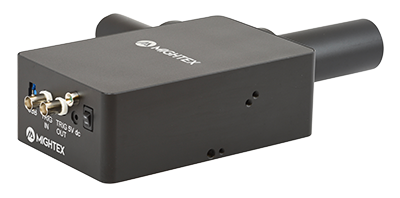
Mightex’s Polygon was used to draw an ROI on the cell to stimulate it with a gradient of blue light intensities for 30 min. This type of control over the intensity and spatial range of illumination would not be possible using other forms of illumination available for confocal microscopy.
Optogenetic Control of Organelle Mobility
Adrian et al. 2017 investigated the interaction between organelles and motor proteins using pattern illumination. They developed a photoswtich optogenetic system based on phytochrome B and activated select areas of the cell with Mightex’s Polygon during live cell imaging.
The Involvement of Erk in Cell Fate Using Optogenetics
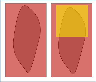
Johnson et al. 2019 investigated how intracellular signaling relates to the development of different cell fates using optogenetics to upregulate Erk activity. Mightex’s Polygon was used to illuminate a section of the cell to eliminate the possibility of global activation leading to found effects.
Spatial Control of Microtubules at the Subcellular Scale
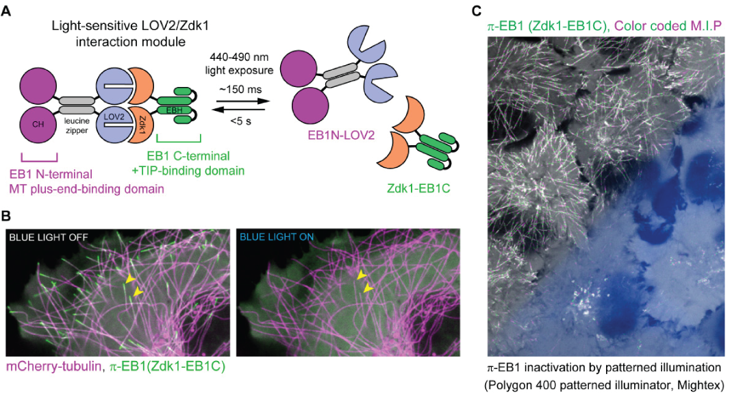
van Haren et al. 2018 illustrated the development of a novel optogenetic system to manipulate microtubule organization. Targeted light stimulation from the Polygon was used to precisely inactivate the microtubule-binding protein End-binding protein 1 (EB1), which is responsible for binding microtubules and recruiting plus-end-tracking proteins (+TIPs) to their positive ends.
Novel Optogenetic System to Control Erk with Spatotemporal Precision
Johnson et al. 2017 investigated the involvement of Erk signaling in embryo development using a novel optogenetic to control the spatiotemporal parameters of Erk activity. Mightex’s Polygon was used to illuminate select portions of the embryo to view differential effects of the optogenetic stimulation on cell development using cell imaging.
Localized Optogenetics
with Mightex’s Polygon
Polygon Inquiry Form:
"*" indicates required fields


