Advances in fluorescent protein technology have made photoactivation techniques broadly available in the cell biology lab. By using these techniques, researchers can fluorescently highlight/label specific protein populations in the cell such that their movement and behavior can be tracked over time. Also, photoactivation methods can be applied at different scales, allowing researchers to track proteins within cells, or alternatively to track cells within tissues.
Localized Photoconversion
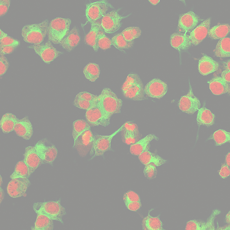
This video shows localized photoconversion of mEos2 to study mitochondrial dynamics in living cells (left) and the results following photoconversion (right)(Courtesy of Dr. Torsten Wittmann from UCSF).
Photoactivation
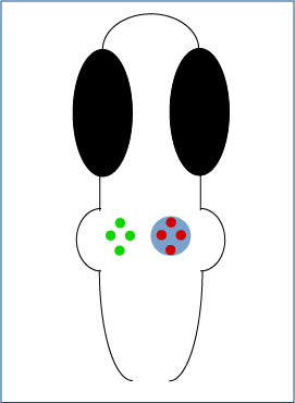
Tabor et al. 2018 investigated in zebrafish the involvement of a subset of neurons in sensory filtering. The zebrafish’s behavioural response was measured in response to ChEF optogenetic stimulation.
Mightex’s Polygon was used to illuminate all neurons expressing ChEF or a subset of these neurons and the behaviour elicited was examined.
Novel Photoactivatable Protein in Zebrafish
Ogasawara 2018 investigated developed a photoactivatable protein to study biological events in zebrafish. Select cells were photoactivated using Mightex’s Polygon.
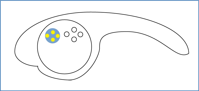
Localized Photoactivation
with Mightex’s Polygon
Polygon Inquiry Form:
"*" indicates required fields


