Understand the Link Between Neural Circuits and Behaviour
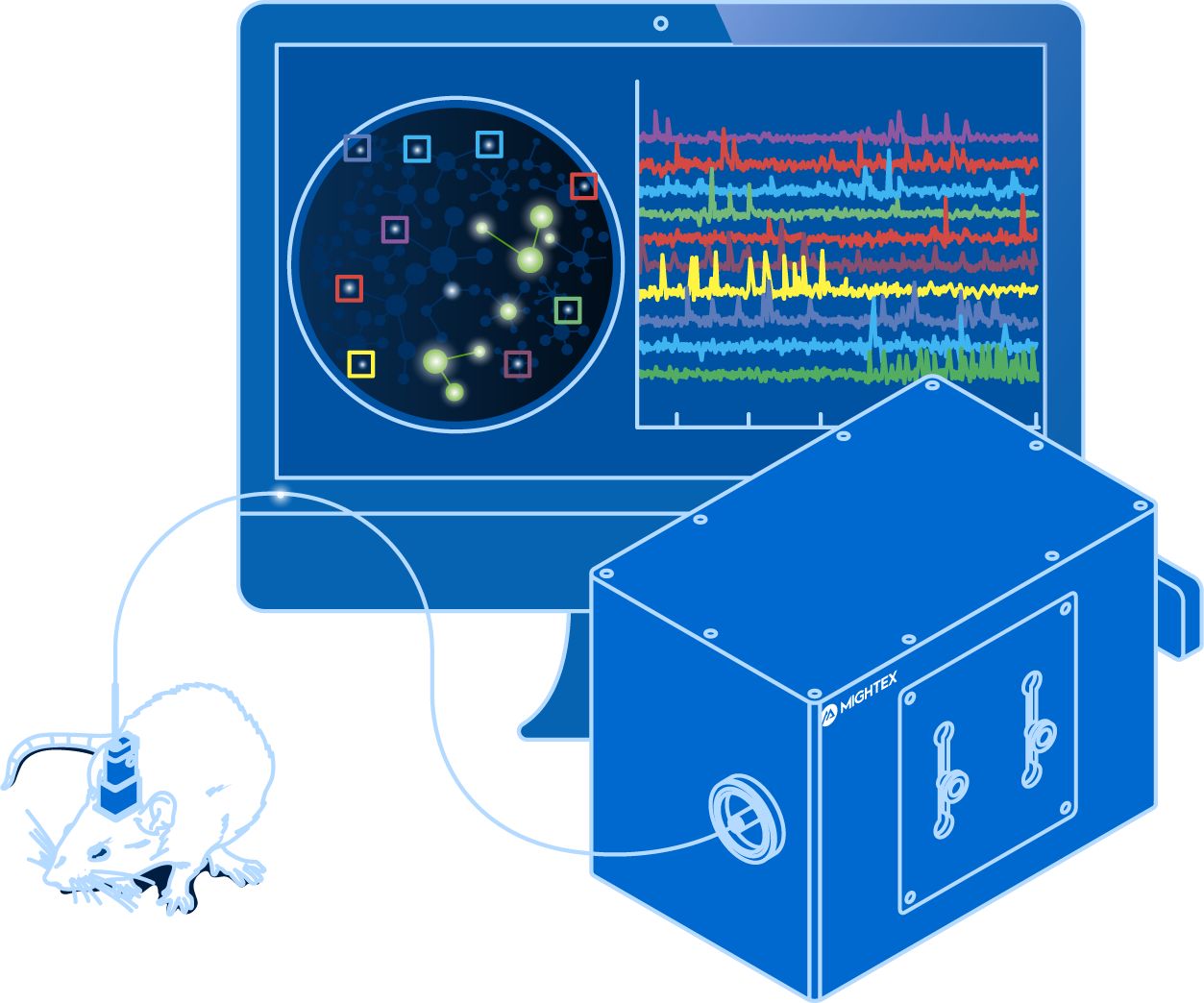
The OASIS Implant is a ground-breaking platform for simultaneous cellular-resolution optogenetics and calcium imaging in freely-behaving animals to probe complex neuronal networks
-
Simultaneous Cellular-Resolution Calcium Imaging and Optogenetics
-
Multi-Region Investigation
-
Reconfigurable Platform
-
High-Quality Imaging with Scientific Cameras
The Evolution in Freely-Behaving Imaging and Optogenetics Technology
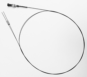
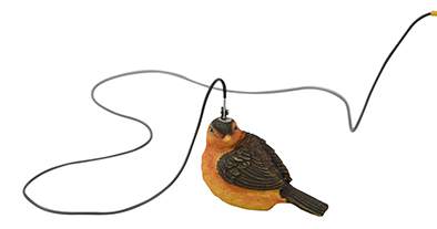
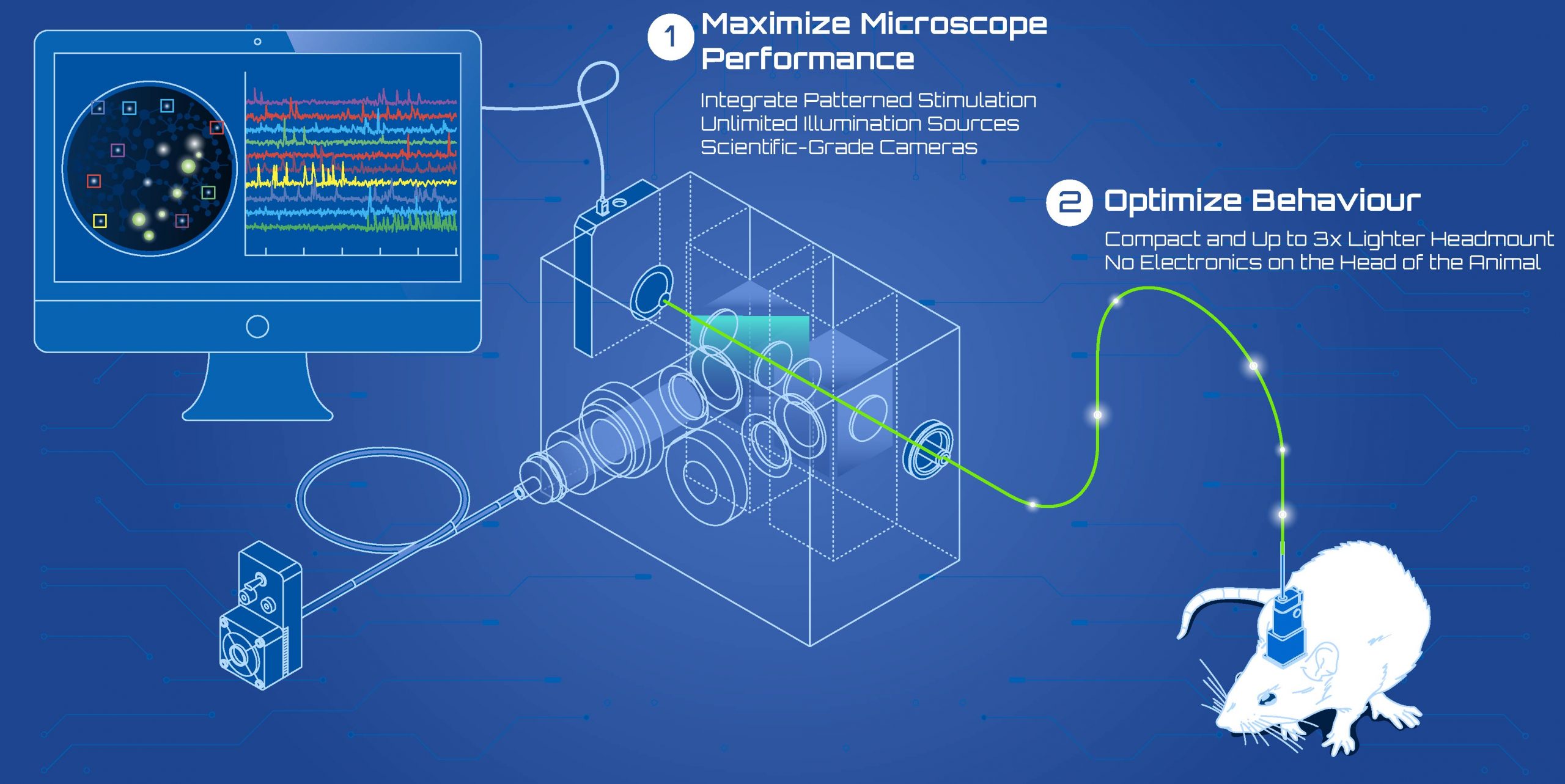
To offer neuroscientists the ability to stimulate and visualize individual neurons in freely-behaving animals, Mightex’s engineers went back to the drawing board and pioneered a new path in the in vivo calcium imaging field. Instead of miniaturizing and placing the microscope on the animal – thereby limiting the microscope’s features and the animal’s behaviour – we kept the microscope off the animal’s head by using an imaging fiber.
By keeping the microscope away from the animal’s head, the OASIS Implant accomplishes 2 main goals:
OASIS Implant Key Features
Investigate Multiple Brain Regions Simultaneously
Simultaneous calcium imaging in the hippocampus (HPC) and prefrontal cortex (PFC) using the OASIS Implant (Courtesy of UCSF).
Neurons project to different parts of the brain, and neuroscientists are interested in understanding how these connections affect behaviour. The OASIS Implant uses a bifurcated imaging fiber that connects to multiple GRIN lenses, allowing researchers to perform imaging and optogenetics in multiple brain regions simultaneously.
Manipulate Functional Neuronal Activity with Cellular-Resolution Optogenetics
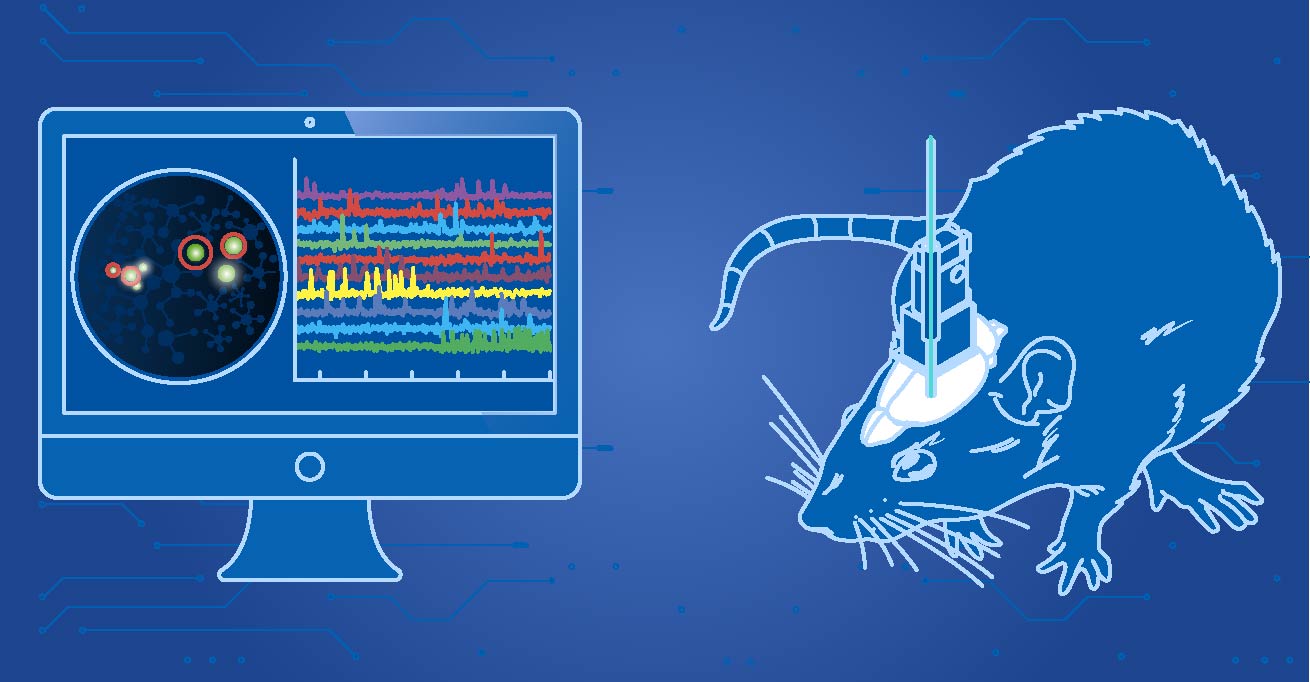
To dissect neural circuits at the level of individual neurons, neuroscientists need a tool to perform cellular-resolution optogenetics in freely-behaving animals. The OASIS Implant uses Mightex’s Polygon DMD Illuminator to illuminate individual neurons in vivo.
Fiber Photometry and Cellular-Resolution Calcium Imaging with One System
GCaMP in vivo calcium imaging in a mouse using the OASIS Implant (Courtesy of Alexxai Kravitz, Washington University in St. Louis).
The OASIS Implant is capable of both population-level calcium imaging using fiber photometry and cellular-resolution calcium imaging. Perform exploratory research using fiber photometry, and delve in deeper to understand the patterns of individual neuron firing.
Micro- to Meso-Scale Imaging and Optogenetics
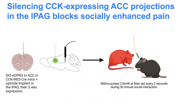
Select optogenetic stimulation of different cortical areas in a freely-behaving animal using the OASIS Implant and Polygon DMD illuminator (Courtesy of Harvard University).
The OASIS Implant enables neuroscientist to perform imaging and optogenetics with cellular-resolution in a deep-region of the brain, or image a large cortical region and stimulate specific populations of neurons (0.5mm to 3.2mm FoV).
Link Neural Activity and Behaviour
GCaMP calcium imaging in a freely-behaving mouse using the OASIS Implant (Courtesy of Alexxai Kravitz, Washington University in St. Louis).
Neuroscientists are interested in linking brain activity and behaviour. Using the OASIS Implant, imaging and optogenetics data can be synchronized with external behaviour cameras and behaviour equipment using the included IO control box to link neural activity and behavioural events.
Compatible with Scientific-Grade Cameras
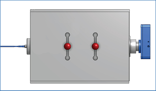
When imaging indicators in vivo, neuroscientists need ways to reliably and confidently collect and analyze the signals obtained. Our platform supports the use of scientific-grade cameras (Photometrics and PCO) that maximize the quality and temporal resolution of the image acquisition. This allows the OASIS Implant to used for applications such as voltage imaging.
Rotation Compensation Option for Freely-Behaving Experiments
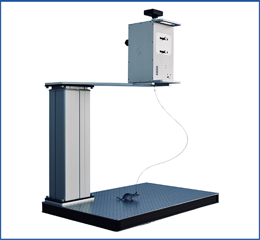
Certain behavioral paradigms require constant animal movement and multiple changes of direction. The OASIS Implant with Rotation Adaptive Mechanism (ROAM) includes cutting-edge technology to allow our imaging fibers to rotate while generating minimal torque on the animal and providing the highest degree of animal movement for studies of naturalistic behaviors.
Reconfigurable System that can be Tailored for Different Experiments
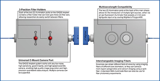
The OASIS Implant is designed to be used for a multitude of imaging and optogenetics experiments. With the ability to easily switch light sources, filter sets, cameras, and fibers, neuroscientists are not limited in the types of experiments they want to perform.
Light-Weight Headmount to Reduce Strain on Animal Behaviour
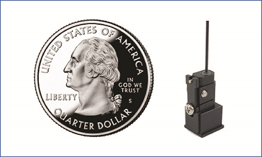
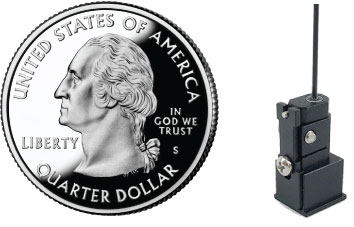
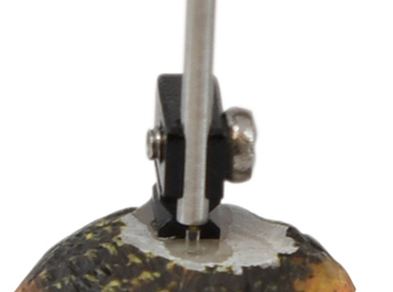
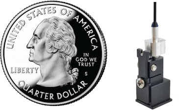
The OASIS Implant uses a light-weight headmount to couple the fiber to the animal’s head for freely-behaving imaging and optogenetics. The headmount (0.7g) is 3 times lighter than a miniscope, and thus, reduces the overall weight on the head of the animal leading to increased animal mobility.
Intuitive Signal Processing for Data Analysis
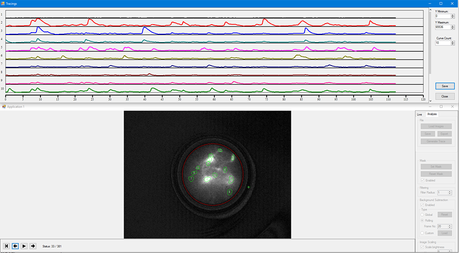
Every OASIS Implant system comes with Mightex’s Image Acquisition and Analysis software that allows users to collect large data sets of neural recordings from calcium imaging experiments and provides the key processing and analysis tools required to help users quickly convert these large data sets into results that can be clearly interpreted for analysis.
1. Acquire In Vivo Calcium Recordings
The software provides a complete set of tools to allow researchers to visualize and capture calcium imaging recordings. The software includes features to synchronize the calcium recordings with the rest of the behavioral equipment and cameras used in the experiment.
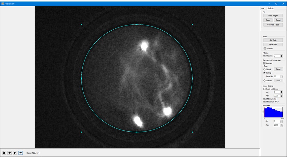
2. Identify Cells
Once calcium recordings are acquired, researchers are able to instantly view and process the captured data. The cell identification tools allow for easy detection of cells and sub-cellular features.
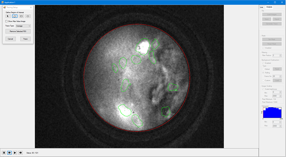
3. Extract DeltaF/F Traces
Traces of the calcium imaging data can be extracted for each cell that is identified. Along with the cell traces, all inputs and outputs to behavioural equipment/cameras will be contained in collected data sets to allow for easy matching of neural events with behavioral activity.
