Published on 2024/12/11 Research powered by Mightex’s Polygon1000 
 Kobayashi, T., Shimba, K., Narumi, T., Asahina, T., Kotani, K., & Jimbo, Y., Revealing single-neuron and network-activity interaction by combining high-density microelectrode array and optogenetics, Nature Communications, 15(1), 9547 (2024).


Introduction
The synchronous activity of neuronal networks plays a crucial role in brain function, yet the interaction between single-neuron activity and network-wide behavior remains poorly understood due to technical limitations in simultaneously stimulating and recording neural activity at the single-cell level. This study explores interactions within cultured networks of rat cortical neurons by integrating high-density microelectrode array recording with targeted optogenetic stimulation using Mightex’s Polygon 1000-G Pattern Illuminator. The approach reveals how individual neurons influence network behavior and how network states, in turn, affect single-neuron responses, advancing our understanding of neural circuit function.
Methods and Results
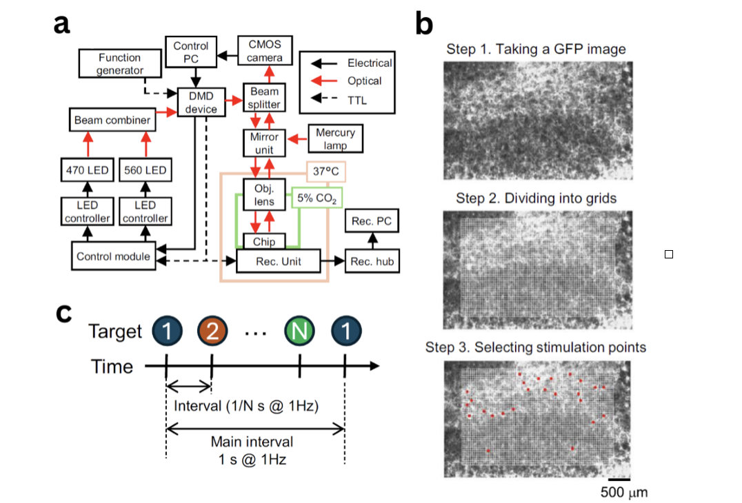
Kobayashi and colleagues developed an experimental setup integrating electrical recording via HD-MEA with optogenetic stimulation. The HD-MEA system contained 26,400 electrodes with a 17.5-μm pitch, enabling high-resolution recording across the neural network. For optogenetic control, they used the Polygon 1000-G system with a spatial resolution of 3 μm, allowing precise targeting of individual neurons (Fig. 1a). The Polygon was controlled with Mightex’s PolyScan software and operated in external trigger mode for precise temporal control of stimulation patterns. The FOV was divided into a grid of 50 × 50 μm² squares and the ones containing ChR2-GFP-expressing neurons were selected and stimulated in random order (Fig. 1b, c).

Figure 1. Experimental setup combining optogenetic stimulation with electrophysiology recordings. a) Schematic showing the optical, electrical and TTL workflows. b) Fluorescence image was taken and divided into grid, grid square locations were selected manually for stimulation. c) Optical stimulus was delivered to each location in sequential order.
When targeting individual neurons with optogenetic stimulation, 77% (244 out of 321) responded within 4.43 milliseconds. The system recorded both directly stimulated neurons and their network-wide effects. The authors identified “leader neurons” that could trigger network bursts with up to 98% probability, and observed that network bursts temporarily altered the response patterns of individual neurons.
Through immunostaining after recording, researchers successfully identified and characterized these leader neurons. The study revealed that leader neurons exhibited intrinsic bursting properties, firing at approximately 100 Hz with 8-second intervals. The combined optical stimulation and HD-MEA recording also enabled detailed mapping of how neural signals propagated from stimulation sites through local circuits, providing insights into network burst generation patterns.
Megha Patwa, Applications Specialist at Mightex
To read the full publication, please click here.
Contact Us
If you want to learn more about using the Polygon in your research get in touch today. Fill out the form below and we will contact you!


