


to optical defocused visual stimuli in mouse retinas
Published on 2024/09/05 Research powered by Mightex’s Polygon1000 ![]()
 So, C., Zhang, T., Wang, Q., Qiu, C., Elie, D. L. A., & Pan, F., The response of retinal ganglion cells to optical defocused visual stimuli in mouse retinas, Experimental Eye Research, 241, 109834 (2024).

Retinal cells capture light and convert it into electrical impulses to be communicated to the brain. It is believed, however, that in addition to this conversion process, they also have the capacity to learn. Indeed, these cells seem to be doing a range of calculations to optimize the image being processed before it even reaches the brain. One important type of calculation involves modifying their output signal in response to blurry light conditions.
Two primary conditions common in humans lead to the blurring of light within the eye: Myopia, otherwise known as short-sightedness, results from the lengthening of the eye, which leads to the lens focusing incoming light in front of the retina rather than on it, and astigmatism results from a misshapen lens which changes the refractive index of the eye.
Fascinatingly, the eye has the capacity to detect the blur of the images that are arriving at its retina and, during developmental periods, change its growth in order to compensate – an ability called ocular compensation (Pan, 2019). While the brain may to some extent be involved in compensating for an unfocused retina, the retina is known to adapt itself. Research in which the eye has been detached from the brain has found that the visual signal being transmitted by the retina is still able to adapt to blurry images (Irving et al., 1992), indicating that a major component of the system in adapting to blurry images is an adaptive retina itself.
In the paper The response of retinal ganglion cells to optical defocused visual stimuli in mouse retinas by So et al, (2024) an array of techniques were used to induce myopic and astigmatic symptoms in retinal tissue. They presented a range of visual stimuli to the myopic and astigmatic tissue and recorded responses from the output cells of the eye, retinal ganglion cells (RGCs), using patch clamping techniques. Together these techniques enabled the researchers to better understand what types of RGCs respond to blurry and focused light and how each subtype responds differently.
Retinal cells were collected from mice, prepared, fixed and patch clamped for electrophysiological recording. These cells, using genetic techniques, were labeled as either ON RGCs or OFF RGCs; ON RGCs depolarize in response to light, while OFF RGCs depolarize in response to darkness.
For the first experiment, an image was projected onto the retinal tissue. To simulate myopia, the image was projected along a range of focus planes, some in front of the retina, some on the retina, and some behind the retina (Figure 1). The researchers found that changes in focus lead to modified activity patterns in 3 of the 4 subtypes of RGCs: both ON and OFF type RGC excitatory post-synaptic currents (EPSCs) were regulated by image focus and OFF type RGC inhibitory post-synaptic currents (IPSCs) were regulated by image focus. These findings confirm that RGCs respond to focused and unfocused light in a variable manner.
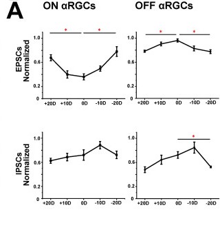
Figure 1: ON and OFF RGC post-synaptic currents in response to different light focus levels. Positive values indicate the image being focused in a plane in front of the tissue; negative values indicate the plane of focus being behind the tissue.
In a follow up experiment, the researchers used a suction pipette to pull on the RGCs, extending them so that they were oblong. This manipulation simulates the effects of myopia, similar to the experiment discussed above. The researchers then illuminated the tissue with images focused in front of, on, and behind the plane of the tissue.The researchers found that the rate of spiking and EPSCs changed in ON-type RGCs in response to stretching (Figure 2). The researchers also found that the rate of spiking and EPSCs changed in OFF-type RGCs in response to stretching. These findings reproduce the work done in the experiment shown in figure 1 while using a more naturalistic method to induce myopia.
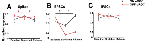
Figure 2: ON and OFF type RGC spiking and post-synaptic currents in response to stretching of the soma.
Finally, in a third experiment designed to replicate astigmatic conditions, bars of light oriented at 4 different angles were projected using a Mightex Polygon1000 digital micromirror system onto retinal tissue. In the control condition, none of the cell types responded differentially to the variable light patterns. In the experimental condition the researchers used a suction pipette to create a negative pressure so as to deform the RGCs in a similar fashion to the experiment described in figure 2. In this condition, the researchers found that OFF RGCs received fewer IPSCs compared to the OFF RGCs in the control condition (Figure 3).
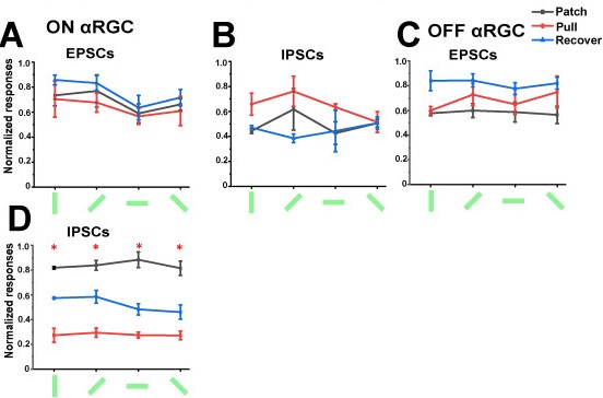
Figure 3: ON and OFF type RGC spiking and post-synaptic currents in response to stretching of the soma and differently oriented light pattern projections.
Altogether, this research supports the previous work indicating that retinal tissue doesn’t simply collect light signals and transform them into electrical signals. The researchers found that, under simulated conditions of both myopia and astigmatism, RGCs acted differently compared to their baseline activity. Given that the activity in these cells is modified by the blurring of light it is clear that retinal tissue can recognize blurring and that this leads to modified RGC activity. It may well be the case that these modified activity patterns are the primary mechanism by which ocular compensation, the change in growth in the eye in response to blurry images arriving at the retina, is triggered.
References
Irving, E. L., Sivak, J. G., & Callender, M. G. (1992). Refractive plasticity of the developing chick eye. Ophthalmic and Physiological Optics, 12(4), 448-456.
Pan, F. (2019). Defocused image changes signaling of ganglion cells in the mouse retina. Cells, 8(7), 640.
So, C., Zhang, T., Wang, Q., Qiu, C., Elie, D. L. A., & Pan, F. (2024). The response of retinal ganglion cells to optical defocused visual stimuli in mouse retinas. Experimental Eye Research, 241, 109834.
Joe Banning, Applications Specialist at Mightex
To read the full publication, please click here.